Nanoparticle tracking analysis characterizes vaccines
01/29/2013
Posted by Lee Mather
Associate Editor, BioOptics World
The virology research group at Lomonosov Moscow State University (MSU; Moscow, Russia) has turned to nanoparticle tracking analysis (NTA) technology for vaccine characterization and standardization, as the technology enables real-time measurement of virus particles in liquid without contamination or particle aggregation (clustering).
The researchers, led by Dr. Nikolai Nikitin, are studying the in-vitro assembly of compositions consisting of artificial plant virus particles and antigens, which are potentially attractive for developing new vaccines. Artificial plant virus particles are spherical particles (SPs) generated by thermal denaturation and structural remodeling of the tobacco mosaic virus (TMV), a rod-shaped virus with a diameter of 18 nm and a modal length of 300 nm. It has been found that upon thermal denaturation of TMV, viral RNA is released and becomes degraded whereas viral-coated protein is assembled into spherical particles. The size of SPs depends on the initial TMV concentration, and particles from 50 to 800 nm may be obtained. The group of Professor Olga Karpova has shown that SPs based on TMV are stable and may adsorb a diversity of proteins. Thus, SPs represent a new type of biogenic nanoplatform attractive for binding antigens and antigenic determinants of different pathogens.
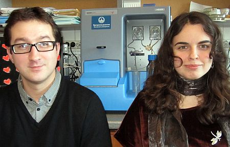 Dr. Nikolai Nikitin and his junior researcher, Ekaterina Trifonova, with the NanoSight NS500 nanoparticle tracking analysis (NTA) system in the Department of Virology at MSU. (Image courtesy of NanoSight/Lomonosov Moscow State University)
The NTA technology (NanoSight's NS500 system), says Nikitin, enabled the research team to analyze and control the size, state of aggregation, and concentration of artificial plant virus particles and small spherical plant and animal viruses. It also allowed them to see the formation of immunogenic complexes (candidate vaccines) by using the indirect immunofluorescence or immunogold staining methods. The technique provided them with the opportunity to obtain simultaneous information concerning nanoparticle size, state of aggregation, concentration, and antigenic specificity in liquid, which are all important for vaccine characterization and standardization, he explains.
Nikitin's research team had previously used transmission electron microscopy (TEM) and dynamic light scattering (DLS) for sizing SPs, isometric viruses, and virus-like particles. To detect the formation of immunogenic complexes (candidate vaccines), they typically use immunogold TEM and immunofluorescence microscopy. However, using NTA instead of these methods eliminates their need to fix and dry the object on a supporting film, which could lead to morphological deformations and aggregation of nanoparticles. NTA also allows analysis of nanoparticles in liquid in real time--something that DLS can also accomplish, but DLS can yield incorrect results due to particle aggregation and sample contamination, Nikitin explains. DLS also cannot estimate the number of particles per unit volume and cannot detect the retention of particles' antigenic properties, but NTA can overcome these limitations by making measurements particle by particle.